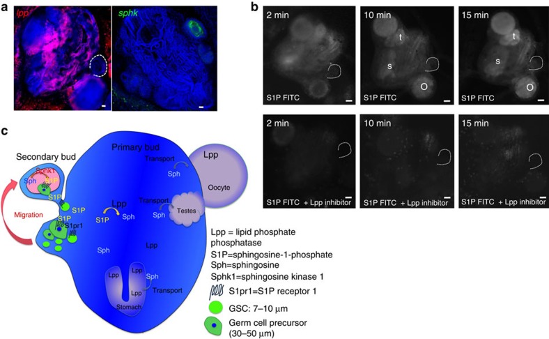
Figure 7. S1P is generated by sphingosine kinase in secondary buds and degraded by LPP in somatic tissues of the primary bud.
(a) FISH for sphingosine kinase (sphk, green) and lipid phosphate phosphatase (lpp, red). Sphk is expressed in double-vesicle-stage secondary buds, whereas lpp is expressed broadly in somatic tissues of the primary bud. Scale bars, 20 μm. (b) Live imaging of fluorescence at different time points after injection of S1P-fluorescein with or without the LPP inhibitor sodium-orthovanadate. After 10 min, S1P-fluorescein accumulates in developing oocytes (o), testes (t) and the stomach (s) of the primary bud. This accumulation is inhibited by inhibition of LPP (n=3). Scale bars, 30 μm (c) Schematic summary of the in vivo data. Vasa-positive germ cells express S1PR1, and migrate towards a gradient of S1P into the germ cell niche in the secondary bud, where S1P is produced by Sphk. In somatic tissues of the primary bud, LPP dephosphorylates S1P to sphingosine, which then gets actively taken up by cells in the developing gonads and the stomach.

