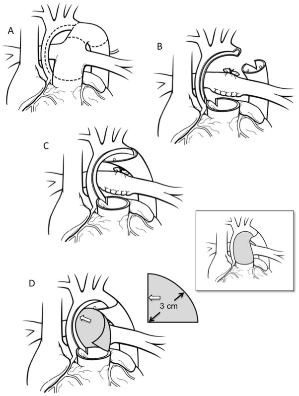
Figure 1.
Coarctectomy combined with an interdigitating arch reconstruction. A) Preoperative anatomy. The dotted lines indicate the areas to be incised. B) The aortic isthmus is divided and all ductal tissue is excised. The undersurface of the arch is incised and this incision is carried down the medial aspect of the ascending aorta. Cutbacks are performed in the posterior left lateral aspect of the proximal descending aorta as well as the pulmonary root leftward of the commissure that is adjacent to the ascending aorta. C) A large interdigitating tissue-to-tissue connection is created between the open distal arch and the descending aorta. The descending aorta can be brought as far proximally as the distal ascending aorta. Finally, the adjacent points of the ascending aorta and pulmonary root cutback are sutured together. D) A patch of homograft material is used to complete the arch reconstruction and neoascending aorta. A flat piece of homograft material is used that is in the shape of a quarter of a circle with a radius of 3cm. The straight edge of the graft (open arrow) is sutured to the inner curvature and the curved outer edge of the patch is sutured to the outer curvature. The patch is tailored as the suture-line transitions to the pulmonary root. Although a large portion of the original patch may be trimmed away the 3cm radius ensures that the neoascending aorta will be without obstruction as this corresponds to the circumference of the typical pulmonary root of the typical patient undergoing the Norwood procedure. The final reconstruction is shown in the inset.

