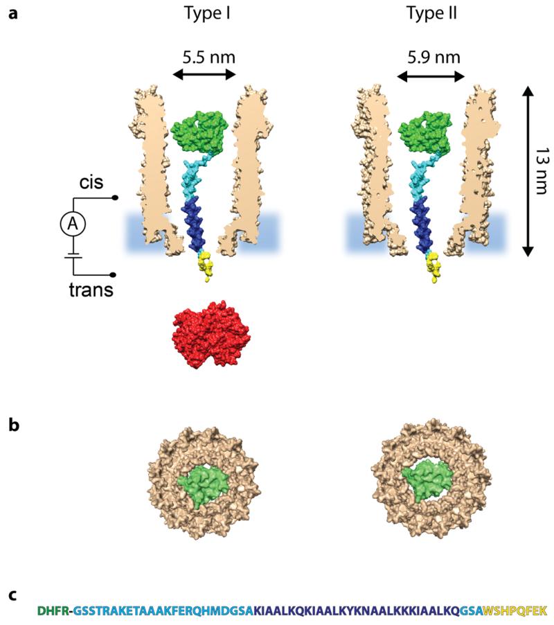
Figure 1. A single DHFRST protein captured inside Type I ClyA-AS and Type II ClyA-CS.
a, Surface representation of a Type I ClyA-AS and a Type II ClyA-CS nanopore (brown, shown as cross section) containing a single E. coli DHFR (green, PDB_ID 1RH3) extended with a polypeptide linker (cyan), a positively charged threading tag (dark blue) and a Strep-Tag (yellow). Strep-Tactin is shown in red. The dimensions of the ClyA nanopore are indicated considering the Van der Waals radii of the atoms.27 b, Bottom view of a single DHFR molecule inside Type I ClyA (left) and Type II ClyA (right), showing the tight fit in dimensions between DHFR and the transmembrane part of Type I ClyA, while in Type II ClyA nanopores DHFR is expected to experience less steric hindrance upon translocation. Both the DHFR and the ClyA nanopore are shown as surface representations. c, Sequence of the DHFR (green) fusion construct, with the polypeptide linker shown in cyan, the positively charged threading tag in dark blue and the Strep-Tag in yellow.

