
Figure 6.
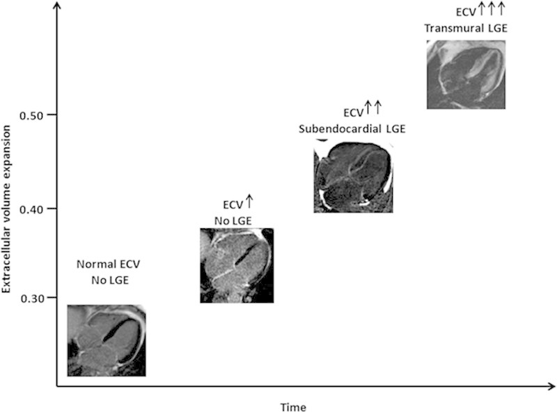
Hypothesized cardiac amyloid progression across time. When amyloid starts to accumulate, 3 steps can be identified: (1) no evidence of late gadolinium enhancement (LGE) but an increase in native T1 and extracellular volume (ECV), (2) a further increase in T1 and ECV and the appearance of subendocardial LGE; and (3) a further increase in native T1 and ECV and progression to transmural LGE.