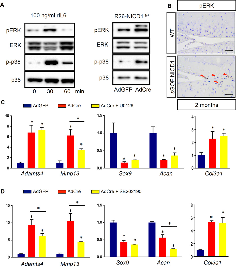
Figure 6. Sustained NOTCH1 signaling activates the ERK/p38 cascade and leads to selected effects on the expression of catabolic, chondrogenic, and fibrous genes.
(A) Western blot analyses for ERK, phosphorylated ERK (pERK), p38, and phosphorylated p38 (p-p38) in proteins extracted from ATDC5 cells treated with rIL6 (100 ng/ml) for 0, 30, and 60 min, and in proteins extracted from control AdGFP infected or AdCre infected R26-NICD1f/+ primary chondrocytes. Results are representative of three independent experiments. (B) IHC analyses of pERK on 2-month-old WT and sGOF NICD1 mutant knee sections. (Scale bars, 50µm.) Red arrowheads indicate pERK positive cells. (C, D) Real-time qPCR comparing gene expression in RNA extracted from control AdGFP infected and AdCre infected R26-NICD1f/+ primary chondrocytes. (C) AdGFP/AdCre infected plus ERK inhibitor U0126, and (D) AdGFP/AdCre infected plus p38 inhibitor SB202190. Gene expression analyses were performed for Adamts4, Mmp13, Sox9, Acan, and Col3a1 in triplicate. All samples were normalized to Beta-actin and then normalized to the controls. Bars represent means ± SD. “*” denotes P<0.05, one-way ANOVA, followed by Bonferroni method.

