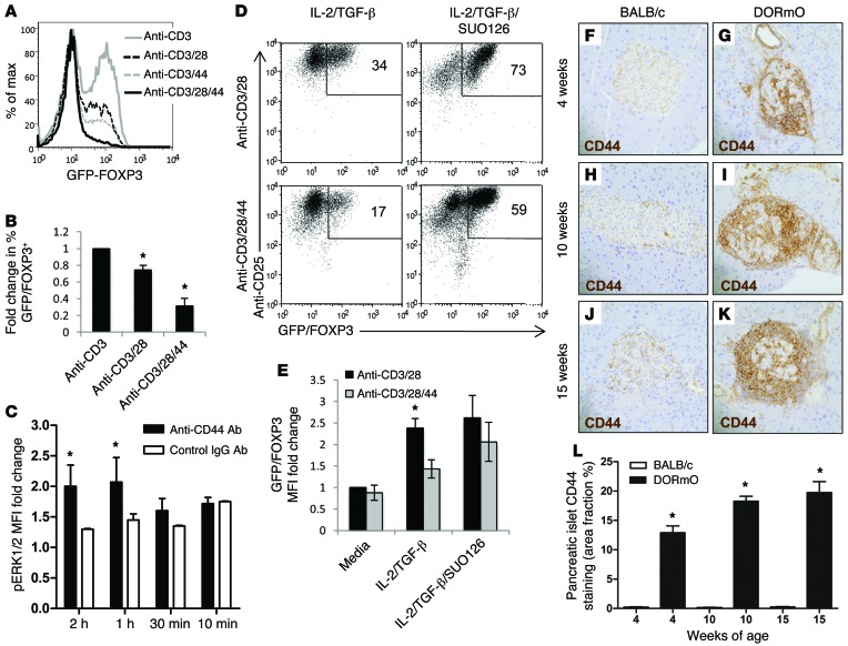
Figure 9. 4-MU treatment promotes FOXP3 induction.
(A) FOXP3 levels following induction from CD4+GFP/FOXP3– T cell precursors performed in the setting of anti-CD3, with or without anti-CD28 and/or anti-CD44 antibody costimulation. (B) Pooled data for 4 independent experimental replicates for the representative data in A. (C) Fold change in pERK1/2 MFI over time following CD44 crosslinking. The data shown incorporate 3 experimental replicates. (D) CD25 and GFP/FOXP3 levels following activation of CD4+GFP/FOXP3– T cells in the setting of TGF-β and IL-2, with or without CD44 costimulation and/or the ERK1/2 inhibitor SUO126. (E) Pooled data for 6 independent experimental replicates for the representative data in D. (F–K) CD44 staining of representative pancreatic tissue sections from BALB/c (control) or DORmO mice fed either 4-MU or control chow. (L) Average CD44+ area of islets for these mice. At least 25 islets were visualized per mouse (n = 6). Original magnification, ×40. Data represent mean ± SEM; *P < 0.05 vs. respective control and control for each time point by unpaired t test.

