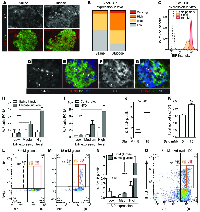
Figure 3. β cells that activate UPR during increased insulin demand are more likely to proliferate.
(A and B) β cell BiP expression was heterogeneous in pancreas sections from glucose-infused mice; hyperglycemia increased the number of β cells with high BiP expression. Representative images are shown from n = 3 (saline) and n = 4 (glucose) mice. (C) In vitro, glucose increased the number of primary mouse islet cells (gated insulin [+]) with high BiP expression. (D–G) In pancreas sections from hyperglycemic mice, PCNA staining frequently occurred in β cells with high BiP expression (arrowheads) and less frequently in β cells with low BiP expression (arrows). (H and I) In pancreas sections from hyperglycemic (H) or high-fat fed (I) mice, β cells containing high BiP were more likely to be PCNA-positive (n = 3–5). (J and K) In vitro, by flow cytometry, glucose increased primary mouse β cell BrdU incorporation and cell number (n = 3). (L–N) Under glucose stimulation, mouse β cells with high BiP were more likely to be BrdU-positive (n = 3). (O) In contrast, in primary mouse β cells overexpressing cyclin D2, BrdU incorporation was not limited to high BiP–expressing cells. (A and D–G) Images acquired at ×200 magnification. Images in D–G and flow plots in C, L, M, and O are representative of n = 3–5 experiments. Data are represented as mean ± SEM; *P < 0.05, **P < 0.01, and ***P < 0.001 by ANOVA (H, I, and N) or Student’s t test (J and K).

