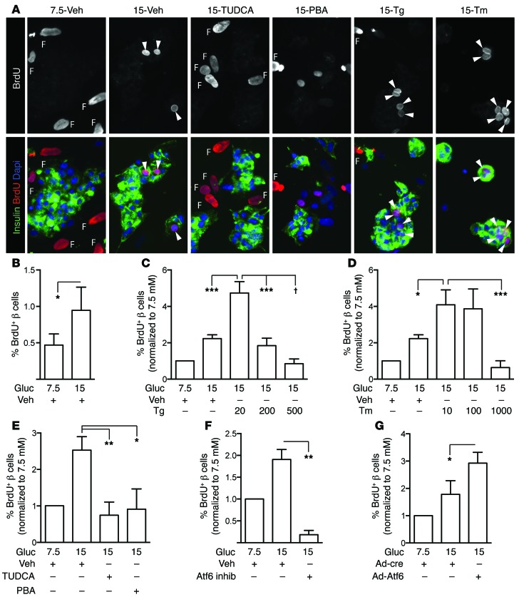
Figure 9. UPR is necessary and sufficient to increase human β cell proliferation, via ATF6.
(A) Human islets were dispersed and cultured on glass coverslips for 96 hours, with BrdU added to culture medium for the entire duration. Arrowheads indicate BrdU-positive β cells. Fibroblast nuclei, which are larger than endocrine nuclei, are labeled F. (B) Human β cell proliferation increased in high glucose (n = 12). Because baseline proliferation varied widely among donors, subsequent panels show data normalized to the 7.5 mM proliferation rate. The raw, nonnormalized data for each prep are in Supplemental Figure 13. (C and D) Low-dose Tg (20 mM, C) or Tm (10–100 ng/ml, D) increased human β cell proliferation (n = 6–12). (E) TUDCA and PBA reduced human β cell proliferation in 15 mM glucose (n = 6). (F and G) In 15 mM glucose, human β cell proliferation decreased in the presence of the ATF6 inhibitor (n = 7; F) and increased with overexpression of ATF6 (n = 4; G). (A) Images acquired at ×200 magnification. Data are represented as mean ± SEM; *P < 0.05, **P < 0.01, ***P < 0.001, and †P < 0.0001 by ANOVA (C–E) or Student’s t test (B, F, and G).

