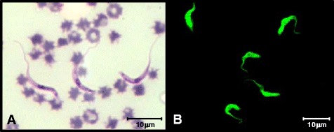
Fig. 1.

a Trypomastigotes of T. vivax in a blood smear of an infected animal dyed with Quick Panoptic and seen using an optical microscope 1,000× magnification. b The form of trypomastigotes marked with fluorescein isothiocyanate from indirect immunofluorescence of a positive animal. Image obtained using confocal microscope (Zeiss® LSM 510 Meta) 1000× magnification
