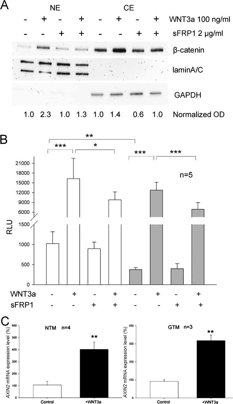
Figure 3.
Existence of a functional canonical WNT signaling pathway in primary HTM cells. (A) Western blot of NE proteins and CE proteins from a primary NTM cell strain treated with or without WNT3a (100 ng/mL) and/or sFRP1 (2 μg/mL) for 4 hours. LaminA/C served as a nuclear protein loading control, while GAPDH served as a cytosolic protein loading control. Three different HTM strains were tested and representative data are shown. The OD of each β-catenin band was normalized first to laminA/C (for nuclear β-catenin) or GAPDH (for cytosolic β-catenin). They then were normalized to corresponding control (non-treated, which was set at 1.0), and the values are shown on the bottom of the Western blot. (B) Lentivirus-based luciferase assays of the canonical Wnt signaling pathway in primary NTM (white columns, left panel) and GTM (grey columns,right panel) cells. HTM cells were transfected with the lentiviral TCF/LEF reporter, and treated with or without 100 ng/mL WNT3a and/or 2 μg/mL sFRP1. Columns represent RLU. Data were analyzed by ANOVA and Bonferroni multiple comparison tests except for the comparison between columns 1 and 5 (from left to right), for which Student's t-test was used. Two different HTM strains were tested and representative data are shown. (C) Comparison of AXIN2 mRNA expression levels of primary NTM (white columns) and GTM (black columns) cells treated without or with 100 ng/mL WNT3a for 4 hours. Left panel: NTM cells. Right panel: GTM cells. Data were analyzed by Student's t-test. Columns and error bars: mean ± SD. **P < 0.010. ***P < 0.001.

