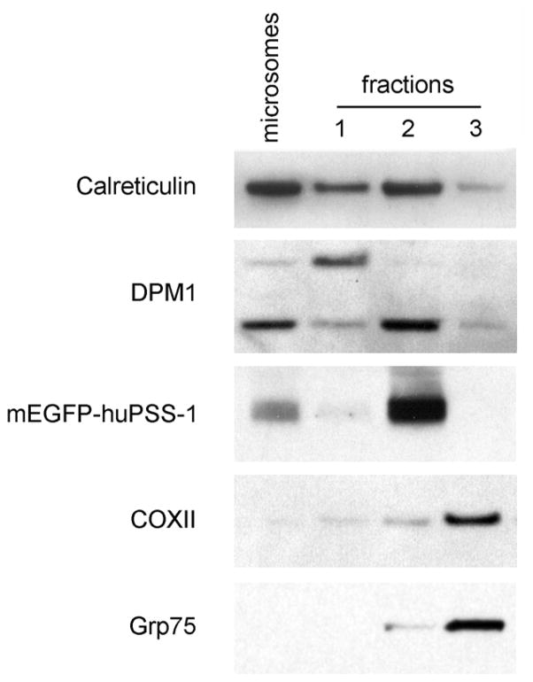
Figure 3.27.6.
Western analyses of fractionated HeLa cells that stably express mEGFP-huPSS-1 fusion protein. Stably transfected cells were fractionated according to the procedure described in Basic Protocol 2 and subcellular fractions were isolated. Fractionated proteins (10 μg) were separated by 10% SDS PAGE and transferred onto nitrocellulose membranes using a semi-dry protein transfer apparatus (BioRad). Blotted proteins were probed against markers for microsomes (anti-DPM1, 1:100 or anti-calreticulin, 1:1000), MAM (mEGFP-huPSS-1 using anti-GFP, 1:100), and mitochondria (anti-COX, 1:100 or anti-Grp75, 1:2500) and with the corresponding horseradish peroxidase-conjugated secondary Ab (1:2000). Reactivity was detected using the chemiluminescent method (Amersham, GE Healthcare).

