
Figure 5. BuGZ coacervation promotes MT polymerization by concentrating tubulin.
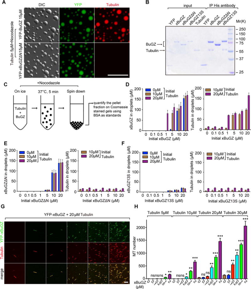
A. Tubulin was concentrated in droplets formed by YFP-xBuGZ, but not by YFP-xBuGZΔN. Scale bar, 20 μm.
B. Anti-His-tag antibody pulled down indicated xBuGZ and tubulin at 4°C.
C. Illustration of the spin down assay.
D. Higher concentrations of YFP-xBuGZ and tubulin are found in droplets (Y-axis) than in initial solution concentrations (X-axis).
E–F. Compared to initial solution concentrations (X-axis), YFP-xBuGZΔN (E) coacervation only concentrated itself, but not tubulin, in droplets (Y-axis), whereas YFP-xBuGZ13S (F) did not coacervate or concentrate itself or tubulin (Y-axis).
G–H. Representative fields of MTs polymerized. MTs were counted in 20 random microscopic fields using a 63× objective. Scale bar, 10 μm.
Error bars, SEM. Student’s t-test: ns, not significant, *p<0.05, **p<0.01, ***p<0.001, three independent experiments.