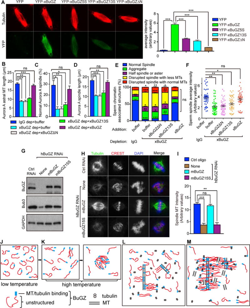
Figure 7. Tubulin/MT binding and coacervation of BuGZ promotes spindle assembly.

A. Incorporation of 0.1 μM YFP-xBuGZ into isolated spindles required Fs and Ys and MT binding. ~30 spindles were analyzed. Scale bars, 10 μm.
B–D. xBuGZ, but not xBuGZΔN or xBuGZ13S, rescued astral MT length (B), percentages of normal spindles (C), and length of spindles (D) induced by Aurora A beads. ~50 (B, D) or ~500 (C) structures were analyzed.
E–F. Only wild-type xBuGZ, but not xBuGZΔN or xBuGZ13S, rescued defective morphology (E) or MT intensity (F) of spindles in xBuGZ-depleted extracts. ~500 sperm-associated MT structures (E) or ~50 spindles (F) were analyzed.
G–I. Expression of mBuGZ, but not mBuGZ15S, in hBuGZ depleted HeLa cells (G) rescued normal spindle MT intensity (H, I). ~30 spindles were analyzed. Spindles, centromeres, and chromosomes were stained. Scale bars, 5 μm.
J–M. Models for BuGZ phase transition in vitro (J–K) or during spindle assembly (L–M). See explanations in the Discussion.
Error bars, SEM. Student’s t-test: ns, not significant, *p<0.05, **p<0.01, ***p<0.001 from three independent experiments. The numbers of structures analyzed are for each experiment and condition.
