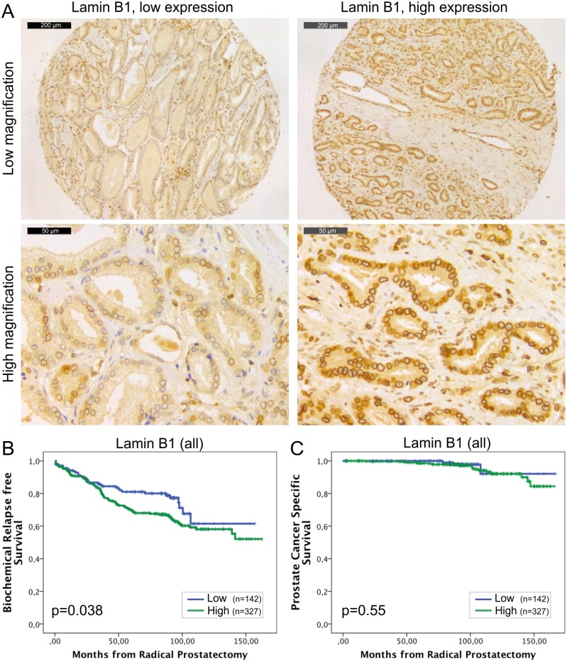
Fig 2. High lamin B1 expression in PCa predicts increased risk for BCR.
(A) Representative examples of TMA slides stained for lamin B1 with immunohistochemistry. Low and high power field images from both low and high expressing tumors are shown. (B-C) Kaplan-Meier analysis indicates that high lamin B1 expression predicts shorter time to BCR in the whole cohort (B; p = 0.038) but has no correlation with DSS (C; p = 0.55).

