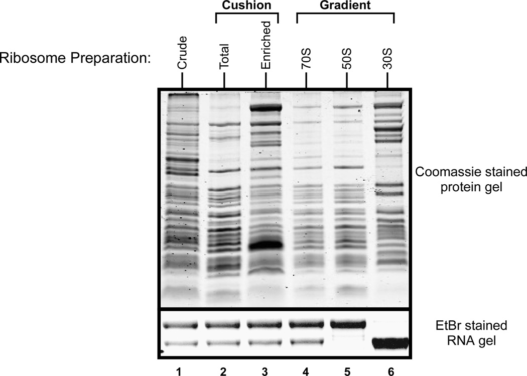
Figure 1. Protein and RNA profile of ribosomes purified by various isolation techniques.
From left to right, Lane 1: Crude Ribosomes, Lane 2: Tight-coupled ribosomes from sucrose cushion, Lane 3: Ribosomes enriched post sucrose cushion, Lanes 4, 5, and 6 are 70S, 50S, and 30S ribosomal profiles, respectively, from an analytical sucrose gradient. Top: Coomassie stained protein gel. Bottom: Ethidium Bromide (EtBr) stained RNA gel showing the 23S and 16S rRNA bands.

