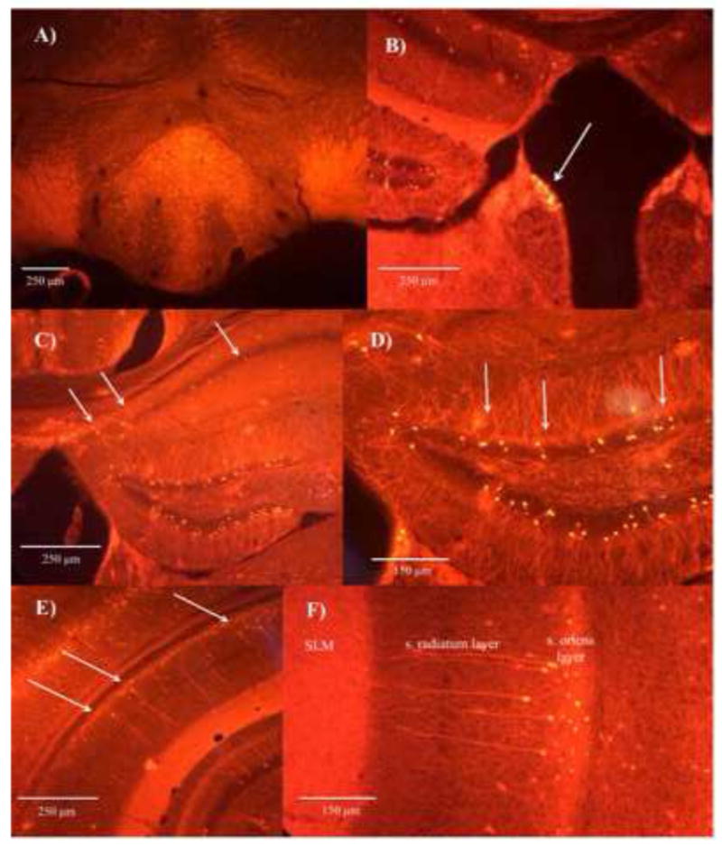
Figure 1. Fluorescent images demonstrate expression of α2-positive neurons.

within the IPN (A), habenula region (B), oriens layer of the dorsal CA1 hippocampal region (C), granular layer of the dentate gyrus (D), and oriens layer of the ventral CA1 hippocampal region (E). The α2-positive neurons within the oriens layer of the CA1 hippocampal region send projections directly into the s. lacunosum moleculare layer (F).
