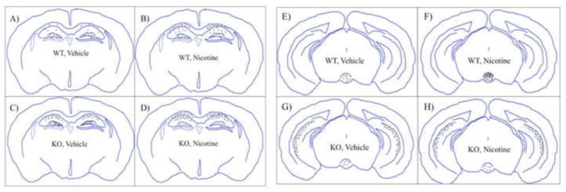
Figure 4. Summary diagram of baseline and nicotine withdrawal-induced neuronal activity across selective midbrain and limbic brain regions.

Representations of cfos activity across the four different groups (A & E. wild type, vehicle; B & F. wild type, nicotine withdrawal; C & G. α2 KO, vehicle; D & H. α2 KO, nicotine withdrawal) within the dorsal hippocampus and dentate gyrus regions (A–D) and the ventral hippocampus and interpeduncular nucleus (E–H).
