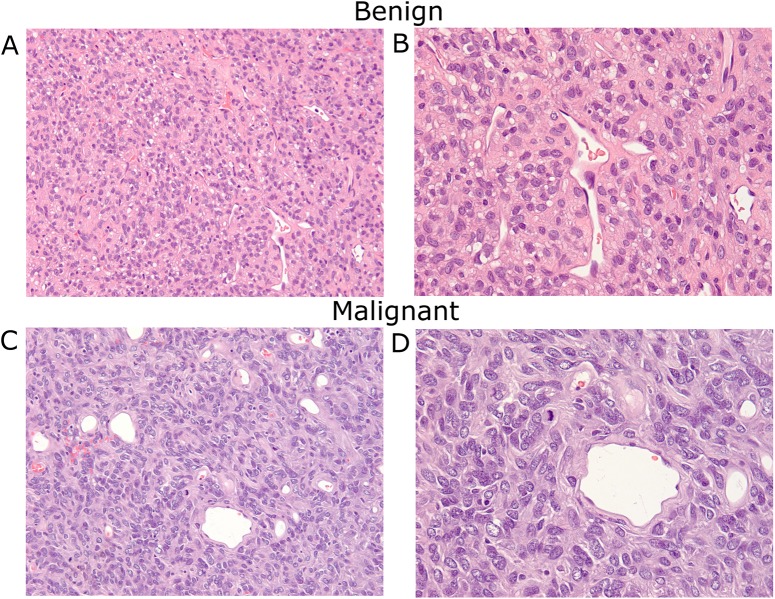
Fig 1. Histologic illustration of benign and malignant SFT.
A: Benign SFT. The tumor shows a patternless architecture composed of spindle cells with hyalinized stroma and thin-walled branching vessels (H&E stain; x200 magnification). B: Benign SFT. The spindle tumor cells have vesicular nuclei without significant cytological atypia, mitosis, and necrosis (H&E stain; x400 magnification). C: Malignant SFT. The tumor has similar architecture; however, cells exhibit marked cytological atypia, including nuclear pleomorphism and increased mitotic activity (H&E stain; x200 magnification). D: Malignant SFT showing atypical mitotic tumor (H&E stain; x400 magnification).

