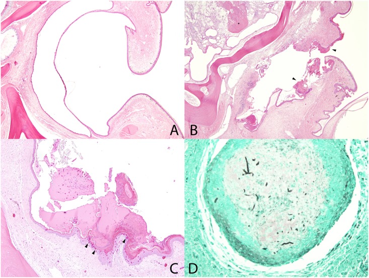
Fig 5. Snake Fungal Disease Histopathology.
Microscopic lesions in cottonmouth snakes inoculated with Ophidiomyces ophiodiicola. A. Normal nasolabial pit. HE stain. B. Nasolabial pit with epithelial thickening and crusts (arrowheads), erosion, ulceration, and dermatitis. Deeper in the head, there is osteomyelitis (asterisk). HE stain. C. Nasolabial pit with crusts and heterophilic dermatitis (arrowheads). HE stain. D. Granuloma deep to the nasolabial pit that contains intralesional fungal hyphae (black linear branching). GMS stain.

