Figure 2.
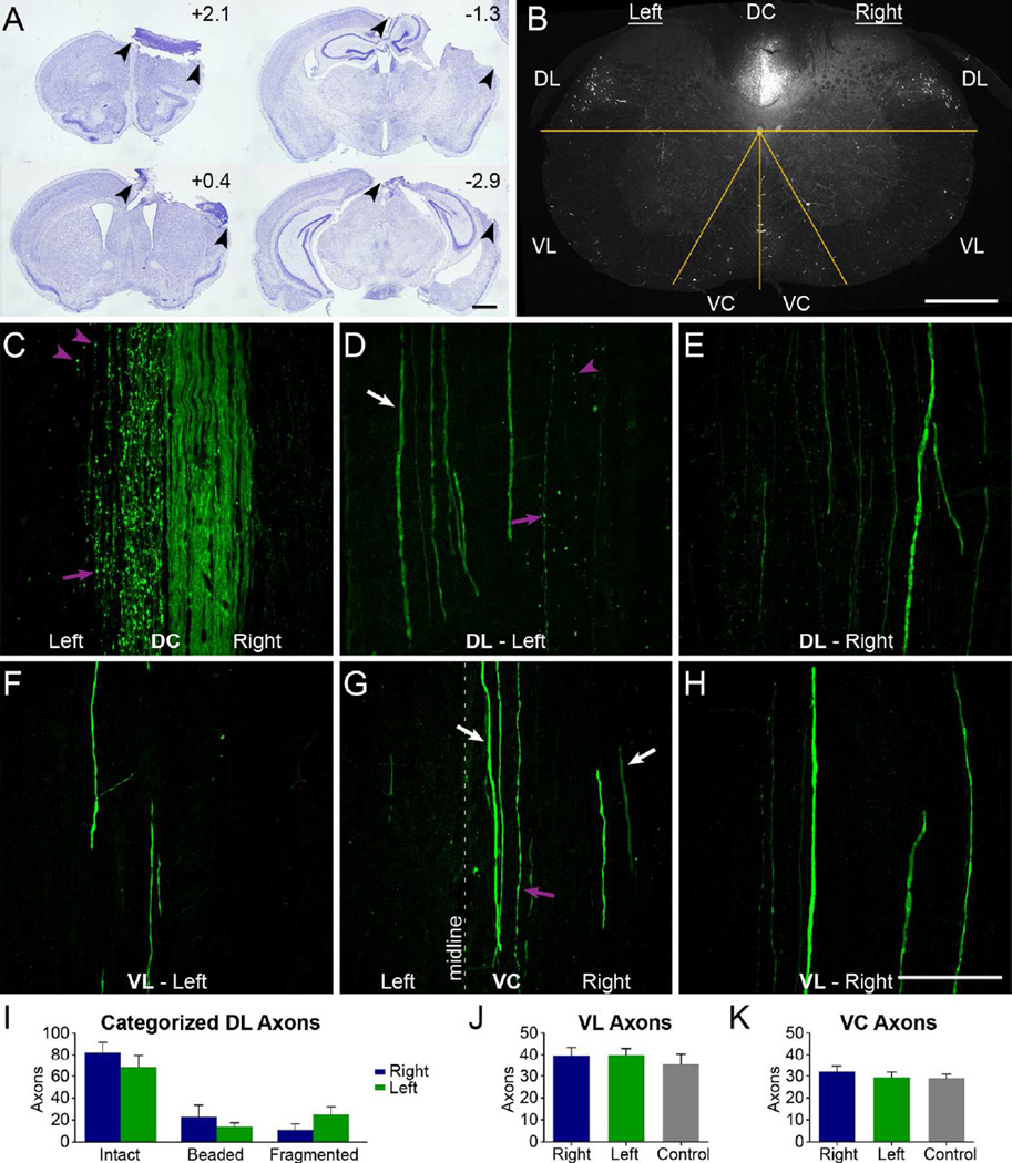
Ablation of the sensorimotor cortex does not lead to degeneration of YFP axons outside the main CST. A, Sections from a representative specimen with a large lesion to ablate CST origins from the right cortex; arrowheads show lesion extent. Rostro-caudal distances of each section from bregma are indicated. B, Confocal cross section of cervical spinal cord 1 week after unilateral cortical ablation. The overlaid yellow lines distinguish the different white matter regions where axons were quantified. C-H, Longitudinal sections of dorsal (C-E) and ventral (F-H) cervical spinal cord, by regions as indicated in B. Purple arrows indicate beaded axons; arrowheads indicate fragmented axons; white arrows indicate intact axons. Note the intact axons in the left DL (D) and right VC (G), as well as bilaterally in the VL (F, H). I, Quantification of categorized axons in the DL in longitudinal sections in mice with a cortical lesion. Differences between sides for the intact, beaded, and fragmented axons were not statistically significant (all p > 0.05, Bonferroni post-hoc tests following 2-way mixed-model ANOVA). J, K, Quantification in cross sections of the overall number of right or left axons in mice with a cortical lesion or axons of control mice in the VL (J) and VC (K), with no respective significant differences (both p>0.6, ANOVAs). The image in C is a single-plane confocal capture and D-H are confocal projections. DC, dorsal column; DL, dorsolateral white matter; VC, ventral column; VL, ventral lateral column. Data are mean + SEM. Scale bars, 1 mm (A), 400 µm (B), 100µm (C-H).

