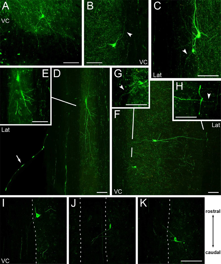
Figure 3.
Some YFP-labeled cells in the spinal cord project into the white matter. A, Cross section of cervical spinal cord showing a YFP-labeled cell in the ventral grey matter with projections extending into the ventral white matter. B-H, Examples of YFP-labeled cells in horizontal sections extending projections into the ventral medial and lateral white matter of the cervical (B, F-H) and thoracic (C-E) spinal cord. Arrowheads highlight some YFP-labeled projections. Each image panel is a projected overlay of 2 adjacent tissue sections except for the images of single tissue sections in inset panels E and G. Inset panels were further adjusted for brightness and contrast to enhance detail. In D, also note the YFP-labeled axon coursing in the ventral root (arrow) and lateral white matter. I-K, Selected confocal projections from mice with large cortical lesions showing YFP-labeled cells in the spinal cord with projections extending both caudally (I-J), and rostrally (K) in the white matter of the ventral column. VC, ventral column; Lat, lateral white matter. Rostral-caudal diagram applies to B-K. Scale bars, 100µm.

