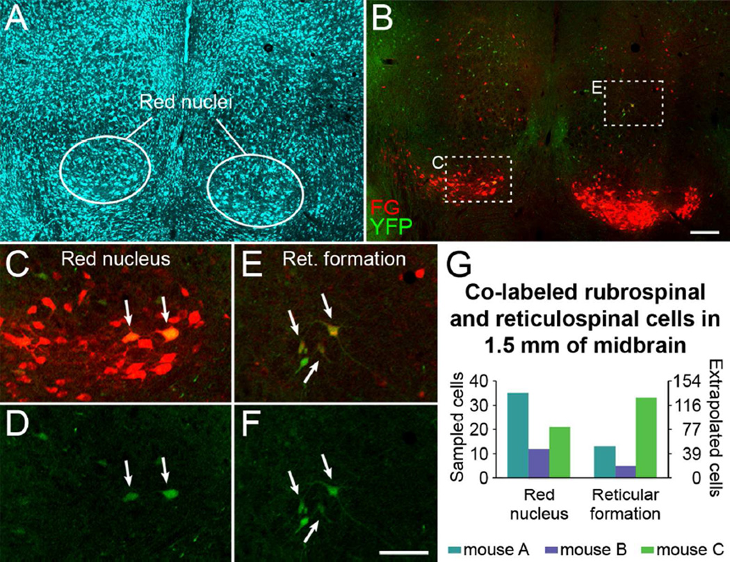
Figure 4.
Some brainstem neurons that project to the spinal cord express YFP. A, Fluorescent Nissl-like labeling (NeuroTrace®) in the midbrain in a mouse that had FG injected into the spinal cord to retrogradely label supraspinal neurons. B, FG and YFP labeling in the midbrain (same section as A). C-F, Merged FG and YFP imaging (C, E) and YFP imaging alone (D, F) in the red nucleus (C, D) and reticular formation (E, F). Note the FG-labeled rubrospinal and reticulospinal neurons that are YFP-positive (arrows). G, Quantification of sampled YFP and FG co-labeled rubrospinal and reticulospinal neurons through 1.5 mm of midbrain in 3 mice. A-F are single plane confocal images. A magenta-green version of this figure is available as Supplementary Figure S2. FG, Fluoro-Gold; Ret. formation, reticular formation. Scale bars, 200 µm (A-B), 100 µm (C-F).

