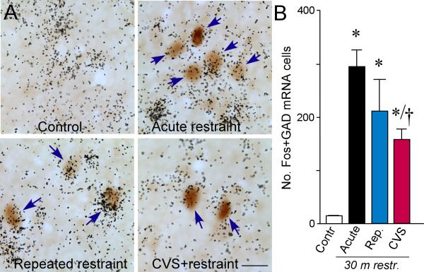
Figure 5.
A: Photomicrographs show representative examples of concurrent labeling for Fos (brown) with GAD67 mRNA (black grains) in doubly-labeled cells (arrows) in aBST (notably within the dorsomedial and fusiform subdivisions). We have previously shown that these cell groups provide a source of GABAergic innervation of CRF-expressing neurons in PVH (Radley et al., 2009; Radley and Sawchenko, 2011). All animals subjected to 30 min of restraint stress showed increases in colabeled GAD+ and Fos+ cells in aBST as compared to unstressed controls, which were void of any Fos expression in this region. CVS resulted in significant decrements in doubly-labeled cells as compared with acutely stressed animals, consistent with the idea that the disinhibition of this pathway may underlie HPA axis sensitization under CVS conditions. B: Mean + SEM number of neurons co-labeled for Fos and GAD67 mRNA in aBST in treatment groups. *, p < 0.05, differs significantly from unstressed controls; †, p < 0.05, differs significantly from the acute stress group. N = 4 control, acute, and repeated groups; N = 5 CVS. Scale bar: 15 μm (applies to all).

