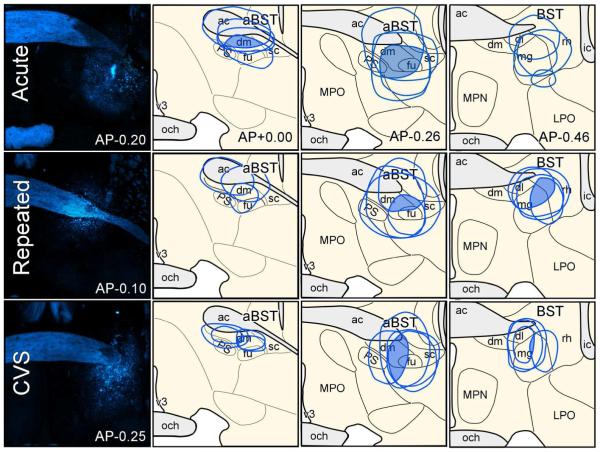
Figure 7.
Reconstructions of Fluoro-Gold (FG) tracer injection placements in aBST in acute, repeated, and CVS rats (top, middle, and bottom rows, respectively). Epifluorescence photomicrographs (left column) depict examples of FG tracer deposits, whereas the shaded regions in the diagrams indicate areas of overlap common to all tracer injections, and their approximate extent of diffusion into adjacent-lying structures. Not shown is a fourth group of unstressed controls were also included in these experiments. ac, anterior commissure; dl, dorsolateral subdivision of aBST; dm, dorsomedial subdivsion of aBST; fu, fusiform subdivision of aBST; ic, internal capsule; LPO, lateral preoptic area; mg, magnocellular subdivision of posterior BST (pBST); MPN, median preoptic nucleus; MPO, medial preoptic area; och, optic chiasm; PS, parastrial nucleus; rh, rhomboid subdivision of pBST; v3, third ventricle.

