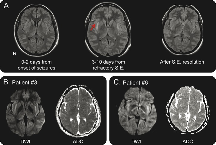
Figure 2. Typical temporal sequence of MRI alterations.
(A) Normal MRI (fluid-attenuated inversion recovery [FLAIR] sequences) before the fully developed status epilepticus (SE) (left); bilateral hyperintensity of the claustrum in FLAIR imaging during status epilepticus (central image); normal appearance of the claustrum after the resolution of the status (right image). Images refer to patient 2. (B, C) Diffusion-weighted imaging (DWI) (left) and apparent diffusion coefficient (ADC) (right) images of patients 3 and 6 show high signal in the region of the claustrum in DWI with normal ADC. The same pattern was observed in other patients. DWI sequences were not acquired in patient 1.

