Figure 3.
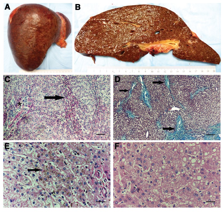
Pathologic findings in the AIP liver. (A) and (B) show gross liver; (C), (E) and (F) are hematoxylin and eosin stains; and (D) shows a Masson trichrome stain. (A) There is a slight nodular appearance of the capsule. No dominant lesions were identified. (B) Cut surface of the explanted liver shows a brown parenchyma with indistinct nodules and minimal fibrosis. (C) Part of a nodule wherein the portal tract is in the center (asterisk) and contains a sprinkling of mature lymphocytes with an accompanying mild ductular reaction. No interface hepatitis is seen. The arrow points to several rows of atrophic hepatocytes that act as the border of the nodule (200×). (D) An area near the hilum showing atrophy of the parenchyma, as evidenced by the approximation of portal tracts. These portal areas, indicated by arrows, are fibrotic with fibrous septa that occasionally bridge and form incomplete nodules. Portal vein branches are obliterated in this photomicrograph (200×). (E) A centrilobular zone showing dark brown pigment granules (lipofuscin) deposited within the cytoplasm of the hepatocytes. Deposition is most prominent in the area indicated by the arrow (400×). (F) Mild steatosis in the form of small and medium droplets is patchy (400×). Scale bars for (C) and (D) = 100 μm and for (E) and (F) = 50 μm.

