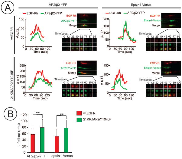
Figure 5. CCP lifetime analysis during internalization of EGF-Rh in HuTu-80 cells expressing wtEGFR or 21KRΔAP2/Y1045F mutant.
wtEGFR and 21KRΔAP2/Y1045F expressing cells were transiently transfected with β2-YFP or epsin1-Venus. After 2 days, time-lapse TIR-FM imaging of cells before and after cell simulation with EGF-Rh (2 ng/ml) was performed as in Figure 4.
(A) Representative examples of fluorescence traces and kymographs from time-series of β2-YFP, epsin1-Venus and EGF-Rh imaging in CCPs which hosted a scission event. A.u.f.i., arbitrary units of fluorescence intensity.
(B) Mean life-time values (+/−S.D.; n=20) of CCPs internalizing EGF-Rh were measured using β2-YFP and epsin1-Venus traces exemplified in (A). **p<0.01.

