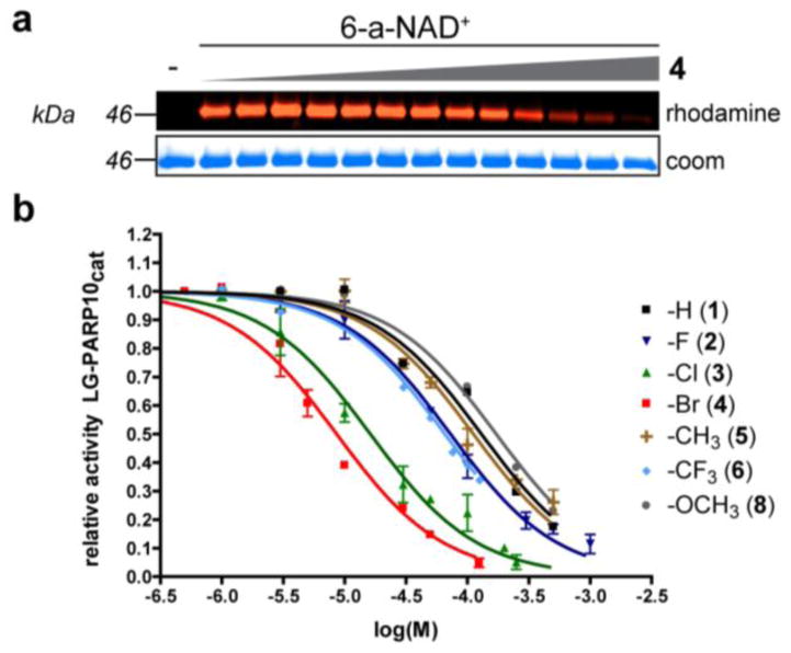
Figure 2.
C-7 substituted dq analogues selectively inhibit LG-PARP10cat. (a) Representative fluorescent gel image showing a dose-dependent inhibition of LG-PARP10cat-mediated ADP-ribosylation of SRPK2 by 7-Br-dq (4). SRPK2 (3 μM) was ADP-ribosylated by LG-PARP10cat (500 nM) with 6-a-NAD+ (100 μM) in the presence of analogue 4 (0 – 125 μM) at 30 °C for 1h. Following click conjugation to rhodamine-azide (100 μM), proteins were resolved by SDS-PAGE and detected via in-gel fluorescence. Coomassie (Coom) Brilliant Blue staining was used to demonstrate even loading. (b) IC50 curves for C7-substituted dq analogues against LG-PARP10cat. Activity was determined as described in (a). Error bars represent S.E.M., n = 2.

