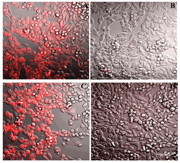
Figure 5.
Fluorescence confocal microscopy. Fluorescent labeling of P2Y14R-CHO cells by 11, analyzed by confocal fluorescence microscopy. Incubation of cells with a fixed concentration of 11 (200 nM) was performed following 30 min (A, B) or 120 min (C, D) of incubation in the absence (A, C) or presence (B, D) of antagonist 5 (10 μM). The complete inhibition of binding was observed in the presence of the antagonist. Scale of 60 μm is shown in D. The excitation laser used had a peak at 555 nm and was equipped with a 561 nm filter. Emitted fluorescence was measured without a filter.

