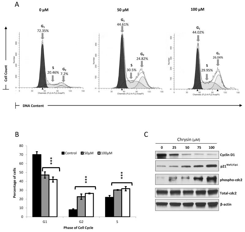
Figure 2. Chrysin causes S/G2 phase arrest in BON cells.
Propidium iodide staining was performed following treatment of BON cells with 0μM, 50μM and 100μM chrysin over a 2 day period. The percentage of cells in the G1, S and G2 phases of the cell cycle in each treated sample was then determined using fluorescent activated cell sorting on the BD FACSCalibur™ platform followed by analysis using the ModFit™ software. Treatment with 50μM and 100μM of chrysin led to an accumulation of cells in the S phase and G2 phase, shown here in one experimental replicate (A). Data from three biological replicates of this experiment were averaged and graphed ± SEM (B) (***p<0.001). Increasing chrysin doses resulted in a decrease in cyclin D1 expression, and an increase in p21Waf1/Cip1 and phosphorylated cdc2, as indicated by Western blotting, thus corroborating the induction of cell-cycle arrest (C).

