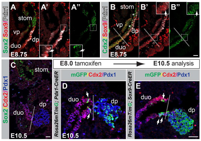
Figure 2. Pdx1 and Sox9 are coexpressed in the pancreatic domain in the foregut endoderm.
(A–B″) Immunofluoresence staining for Sox2, Sox9 and Pdx1 (A–A″) and Cdx2, Sox9 and Pdx1 (B–B″) on embryonic sections at embryonic day (E) 8.75. The arrows in A′,A″ and B′,B″ indicate Pdx1+/Sox9+ cells co-expressing Sox2 and Cdx2, respectively. The dashed line in B–B″ demarcates the transition from the presumptive duodenal to the pre-pancreatic region. Fields demarcated by white dashed boxes in A′,A″,B′,B″ are shown at higher magnification in the same panels. Non-specific signal for Cdx2 is evident in the foregut lumen (B,B″, asterisks) due to antibody trapping. (C) Immunofluoresence staining for Cdx2, Sox2, and Pdx1 at E10.5. (D,E) Dams carrying R26mT/mG embryos expressing CreER driven by either the Pdx1 or Sox9 regulatory sequences were injected with tamoxifen at E8.0, embryos sectioned at E10.5, and immunofluorescence staining performed for Cdx2, Pdx1 and GFP. Recombined, membrane-targeted GFP+ (mGFP+) cells trace to the pancreatic epithelium; scattered labeled cells are also detectable in the proximal duodenum in R26mT/mG;Pdx1-CreER (D) and R26mT/mG;Sox9-CreER (E) embryos. dp, dorsal pancreas; vp, ventral pancreas; duo, duodenum; stom, stomach. Scale bars = 50 μm (A–E).

