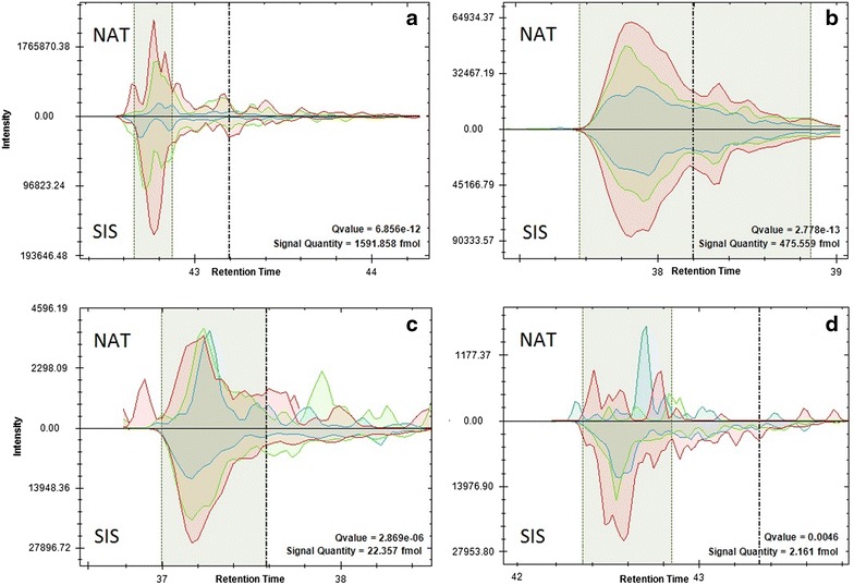
Fig. 1.

Extracted ion chromatograms of native and isotopically-labeled proteotypic peptides (marked as “NAT” and “SIS”, respectively) visualized with SpectroDive™ software for qualitative and quantitative analysis using three transitions per peptide: a SVLGQLGITK (Alpha 1-antitrypsin, P01009), high concentration; b GGYTLVSGYPK (Hemopexin, P02790), moderate concentration; c GYTQQLAFR (Complement C3, P01024), low concentration; d AVSPLPYLR (von Willebrand factor, P04275), in traces. Shaded zones indicate peak integration boundaries. Dashed lines mark peptide elution time predicted by iRT-calibration
