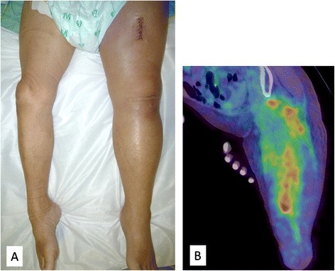
Fig. 1.

Photograph and positron emission tomography-computed tomography (PET-CT) image of the left femur. a Swelling of the left femur. An open muscle biopsy was performed. b PET-CT scan image showing increased metabolic activity in multiple skeletal muscles (latissimus dorsi, transverse, iliac, iliopsoas, and femoral)
