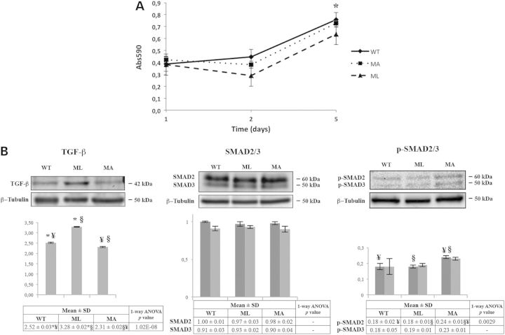
Figure 4.
Osteoblast proliferation and expression analysis of TGF-β and its effector SMAD2/3 in bone from MA, ML and WT mice. (A) Proliferation in calvarial osteoblasts from ML mice is significantly impaired at d5 from plating (*P < 0.05). Each point was measured in triplicate, and each assay was repeated in three independent experiments. The mean values ± SD are reported. (B) Representative western blots using TGF-β antibody (left panel), SMAD2/3 antibody (center panel) and p-SMAD2/3 antibody (right panel). The densitometric analyses obtained by multiple replicative experiments are reported below western blotting images. TGF-β is up-regulated in ML mice whereas p-SMAD2/3 is up-regulated in MA animals. Histograms visualize normalized mean relative-integrated density ± SD values. *, ¥ and § symbols indicate the significance of expression changes occurring between WT and ML, WT and MA, and ML and MA mice, respectively. In SMAD2/3 histogram, dark gray bars represent SMAD2 and light gray bars represent SMAD3; in p-SMAD2/3 histogram, dark gray bars represent p-SMAD2 and light gray bars represent p-SMAD3.

