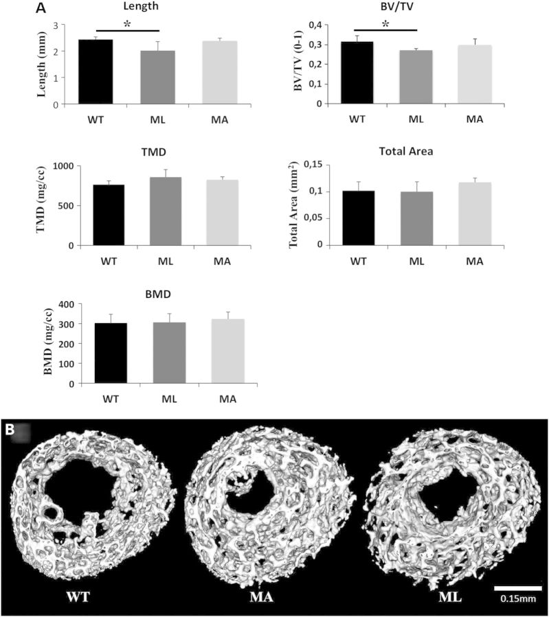
Figure 6.
NanoCT was used to assess mineralized bone parameters in 1-day-old mouse femurs. (A) Lethal Brtl+/− mice showed a reduction in mineralized length between proximal and distal secondary ossification centers, and a reduced bone volume fraction of the trabeculated structure (BV/TV). TMD remained unchanged, and no significant differences in total cross sectional area and BMD were observed between genotypes. (E) Representative nanoCT images of femoral cross sections show a highly trabecular architecture.

