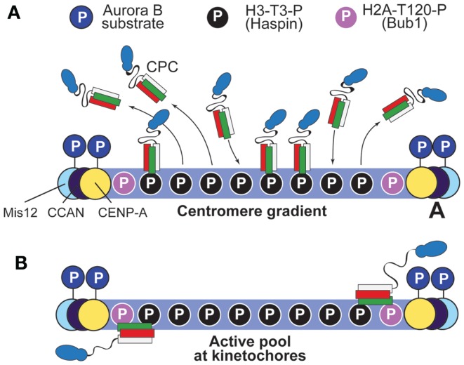
Figure 6.
Two possible models to account for the gradient of Aurora B phosphorylation. (A) The “centromere gradient” model predicts that the CPC becomes recruited to centromeres as illustrated in Figure 4B, and it then dissociates from them, creating a gradient of CPC concentration (and therefore, by inference, of substrate phosphorylation). We note, however, that it is unlikely that this gradient could account for the sharp transition of phosphorylation potential of Aurora B within the very limited scale length of the kinetochore (see Figure 5). (B) An alternative model posits that an active form of the CPC is anchored near the kinetochore, and that the centromere pool is not strictly required for function (it was therefore omitted from the drawing). Proximity to H2A-T120-P might lead to the activation of this kinetochore pool of the CPC. Interactions with kinetochore subunits are also possible.

