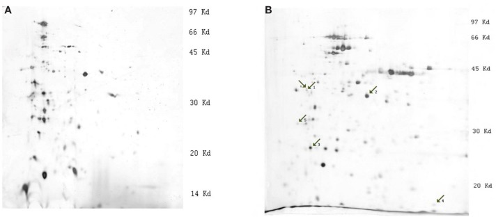
Figure 1.
Images of silver stained 2-D gels of Y. pestis protein extracts (strain 1853, condition 28°C). (A) 2-D electrophoresis on strips with pH linear gradient 3.0–10.0, (B) 2-D electrophoresis on strips with pH linear gradient 3.0–5.6. The numbered arrows indicate the positions of differentially expressed proteins: 1, outer membrane protein C, porin; 2, outer membrane protein C2 porin; 3, Tellurium-resistance protein; 4, DNA-binding protein H-NS; and 5, F1 capsule antigen.

