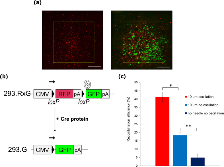
Figure 5. Delivery of Cre recombinase into live cells using the nanoneedle array.
(a) Fluorescent images of 293.RxG cells transfected with the Cre protein with use of 10 μm pitch nanoneedle array after 1 min (left) and 48 h (right). Following successful insertion, cells that underwent recombination due to Cre delivery express GFP instead of RFP, showing in green color. Yellow square of 3 × 3 mm represents the contact area covered with the nanoneedle array. Scale bars are 1 mm. (b) Structure of the reporter gene in 293.RxG cells before and after recombination with Cre protein. (c) Recombination efficiency with Cre protein delivered by 10 μm pitch nanoneedle array with or without oscillation and with use of a flat silicon wafer (n = 3). Two-sided Student’s t-test was performed. *P = 0.0026, **P = 0.0021. Error bars are SD.

