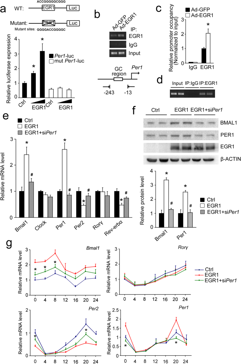
Figure 6. PER1 mediates the regulation of clock genes by EGR1.
(a) Reporter gene assays in 293T cells using wild-type (Per1-luc) or EGR1 binding site mutant (mut Per1-luc) Per1-luciferase reporters in combination with EGR1 or control plasmid. *P < 0.01, EGR1 versus control. (b) ChIP assays with the indicated antibodies using AML-12 cells infected with GFP or EGR1 adenoviruses for 48 h. PCR primers amplified a fragment flanking the GC region on the Per1 promoter. (c) qPCR analysis of ChIP assays with the indicated antibodies. *P < 0.01. (d) ChIP assays in mouse livers at ZT13. PCR primers amplify a fragment flanking GC region on the Per1 promoter. (e) qPCR analysis of clock genes in primary hepatocytes infected with the GFP or EGR1 adenoviruses for 48 h after transfection with Scrb or Per1 siRNA for 6 h. *P < 0.04, EGR1 versus control. #P < 0.05, EGR1+siPer1 versus EGR1. (f) Protein expression in primary hepatocytes infected with the GFP or EGR1 adenoviruses for 48 h after transfection with Scrb or Per1 siRNA for 6 h. The gels were run under the same experimental conditions. The full-length blots are presented in Supplementary Fig. 8. *P < 0.02 EGR1 versus control. #P < 0.02, EGR1+siPer1 versus EGR1. (g) Time course expression of clock genes in mouse AML-12 cells infected with GFP or EGR1 adenoviruses for 48 h after transfection with Scrb or Per1 siRNA for 6 h following serum shock. *P < 0.05 EGR1+siPer1 versus EGR1.

