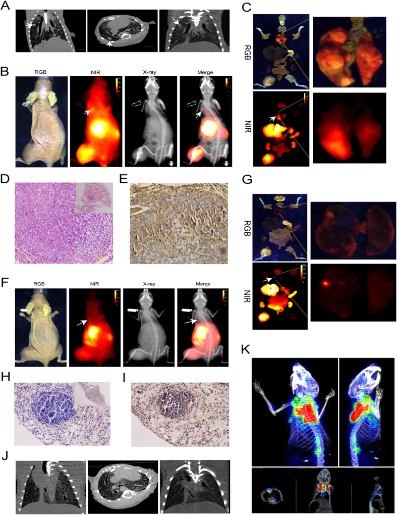
Figure 3. CXCR4-IR-783 allows detection of lung metastasis of osteosarcoma.
(A) Micro -CT scan shows the presence of metastatic lesions (arrows) in the lungs of mice bearing F5M2 xenografts 6 weeks after inoculation. (B) The metastasis in the lungs is visualized by injection of CXCR4–IR-783 6 weeks after inoculation of F5M2 cells. The NIR image shows the presence of an apparent signal (arrow) in the lung, which is confirmed by the merged image of NIR and X-ray images. (C) RGB (upper panel) and NIR (lower panel) signal of all organs (left panel) and dissected lung tissue (right panel) of mouse bearing F5M2 cells 6 weeks after inoculation. (D) H&E staining reveals the presence of osteosarcoma cells in the lung tissues (400 × and 40 × (inset)). (E) Immunohistochemistry shows apparent CXCR4 expression in the metastatic osteosarcoma tissue (400 × ). (F) NIR fluorescence signal (arrow) is detected in the lungs of mouse bearing F5M2 xenografts 4 weeks post tumor implantation. (G) The RGB (upper panel) or NIR (lower panel) images of all ex-vivo organs (left panel) and lung tissue (right panel). The presence of osteosarcoma metastasis in the lung tissues is confirmed by histopathologic evaluation with H&E staining (H, 400 × and 40 × (inset)) and by immunohistochemistry for CXCR4 (I, 400 × ). The metastatic lesions in the lungs could be detected using Micro-CT scan (J) and 18F-FDT PET scan (K) of the chest failed to reveal the high glucose uptake of the nodules in the lungs.

