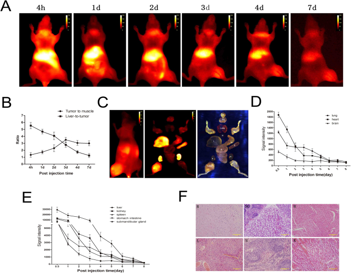
Figure 4. CXCR4-IR-783 exhibits time-dependent clearance from normal mouse organs.
(A) The temporal distribution of CXCR4-IF-783 in mice bearing F5M2 osteosarcoma xenografts three weeks after tumor implantation by contiguous NIR imaging. Animals are shown in the ventral view. (B) The tumor-to-muscle (pellets) and liver-to-tumor ratio (squares) ratios following injection of CXCR4-IF-783. Data shown are mean ± SD. N = 10. (C) The NIR image in vivo (left) and ex vivo (middle), and the RGB image show the corresponding dissected organs (right). Contiguous NIR imaging demonstrates a steady decline in the mean NIR fluorescence intensity in the lungs, heart and brain (D) and the liver, spleen and other organs as well as the tumor xenograft from the day of injection of CXCR4-IR-783 to day 8 post injection (E). (F) H&E staining confirms the tissue type in the organs. B, brain, SG, submandibular gland, H, heart, L, liver, S, spleen and K, kidney (K) (400 × ).

