
Figure 8.
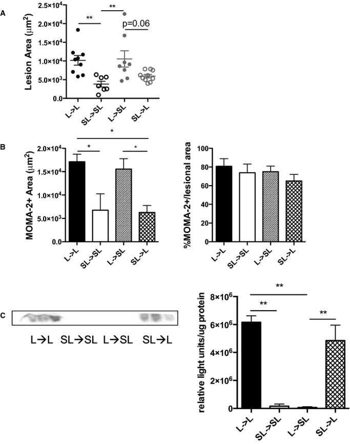
SAA produced by macrophages may promote atherosclerosis and increase the amount of lesional macrophages. Bone marrow transfer recipients were fed a Western-type diet for 6 weeks. A, The ascending aorta was stained with oil red O, and cross-sectional area was quantified. Each dot represents an individual mouse. n=7 to 10 for all groups. B, MOMA-2+ lesional area was quantified in the ascending aorta in chimeric Ldlr−/− and Saa−/−Ldlr−/− recipient mice. n= 4 to 5 per group. C, Plasma SAA levels in chimeric Ldlr−/− and Saa−/−Ldlr−/− recipient mice were analyzed by Western blot. Data are expressed as relative light units per microgram of plasma. n=3 per group. *P<0.05; **P<0.01. L indicates Ldlr−/−; SAA, serum amyloid A; SL,Saa−/−Ldlr−/−.