
Figure 10.
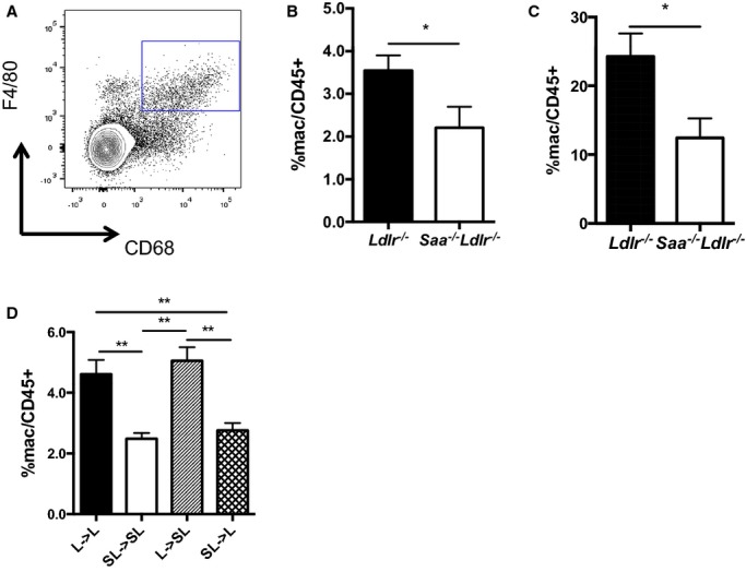
The absence of SAA produced by macrophages reduces macrophages in the spleen in Ldlr−/− mice fed a WTD. A, Macrophages in the spleen were quantified using flow cytometry and defined as CD45.2+F4/80+CD68+. Percentage of macrophages in the (B) spleen and (C) peritoneum of Saa−/−Ldlr−/− and Ldlr−/− mice fed a WTD for 6 weeks. Data are compiled from 2 pooled experiments for each group. n=8 to 10 per group. D, Percentage of macrophages in the spleen of Saa−/−Ldlr−/− and Ldlr−/− chimeric mice fed a WTD for 6 weeks. n=7 to 9 per group. Representative of 2 pooled experiments for each group. Percentages were normalized to CD45.2+ cells. *P<0.05, **P<0.01. L indicates Ldlr−/−; SAA, serum amyloid A; SL, Saa−/−Ldlr−/−; WTD, Western-type diet.