Figure 5.
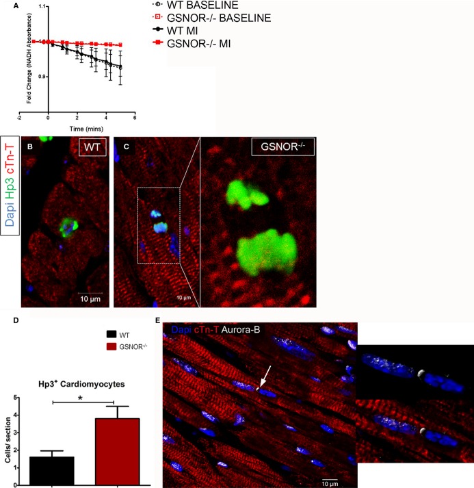
Enhanced mitotic activity of cardiomyocytes in GSNOR−⁄− hearts post MI. A, Absorption of NADH in whole-heart lysates from GSNOR−⁄− and WT mice, before and 8 weeks after the occurrence of MI. No NADH oxidation is recorded before the addition of GSNO in WT or GSNOR−⁄− lysates (time −2 to 0). Addition of GSNO (time 0), produces a sharp decrease in the absorbance of NADH in WT (black lines), but not in GSNOR−⁄− (red lines) heart lysates, before and after MI. These results indicate that the adult myocardium is enriched in GSNOR activity before and after MI, and that GSNOR activity is diminished in the pre- and post-MI GSNOR−⁄− heart. B and C, Confocal immunofluorescence analysis of cardiomyocyte mitosis, based on the nuclear expression of Hp3, in WT (B) and GSNOR−⁄− (C) hearts, 1 week post MI. D, Quantification of Hp3+ cardiomyocytes reveals that, compared with WT,GSNOR−⁄− hearts are characterized by a 3-fold increase in cardiomyocyte mitosis, 1 week post MI (t test; *P= 0.01). E, Representative confocal immunofluorescence image illustrating expression of Aurora-B kinase, a marker of cytokinesis (arrow), at the cleavage furrow of a mitotically dividing GSNOR−⁄− cardiomyocyte, 1 week post MI. Values are mean±SEM. cTn-T indicates cardiac troponin T; GSNOR, S-nitrosoglutathione reductase; MI, myocardial infarction; NADH, reduced form of nicotinamide adenine dinucleotide (NAD); WT, wild-type.

