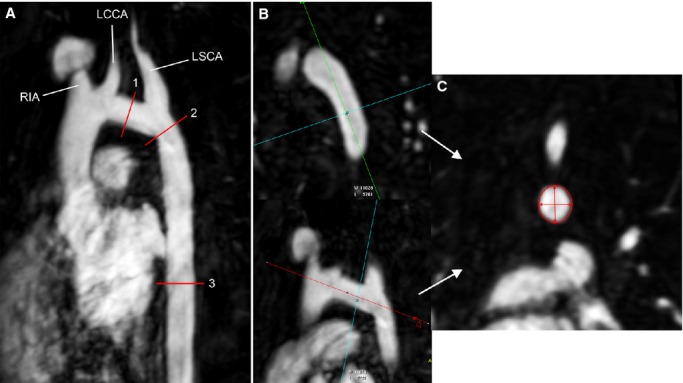
Figure 1.

A, Maximum intensity projection from the gadolinium 3-dimensional (3D) MRA showing the sites of measurements. The transverse aortic arch (1) extending from the origin of the RIA to the origin of the LSCA was measured at its narrowest segment. The isthmus (2) was measured just distal to the origin of the LSCA. The thoracic descending aorta (3) was measured at the level of the left atrium. B and C, Example of multiplanar reformatting of the 3D MRA to measure 2 orthogonal diameters and cross-sectional area of the transverse aortic arch. LCCA indicates left common carotid artery; LSCA, left subclavian artery; MRA, magnetic resonance angiogram; RIA, right innominate artery.
