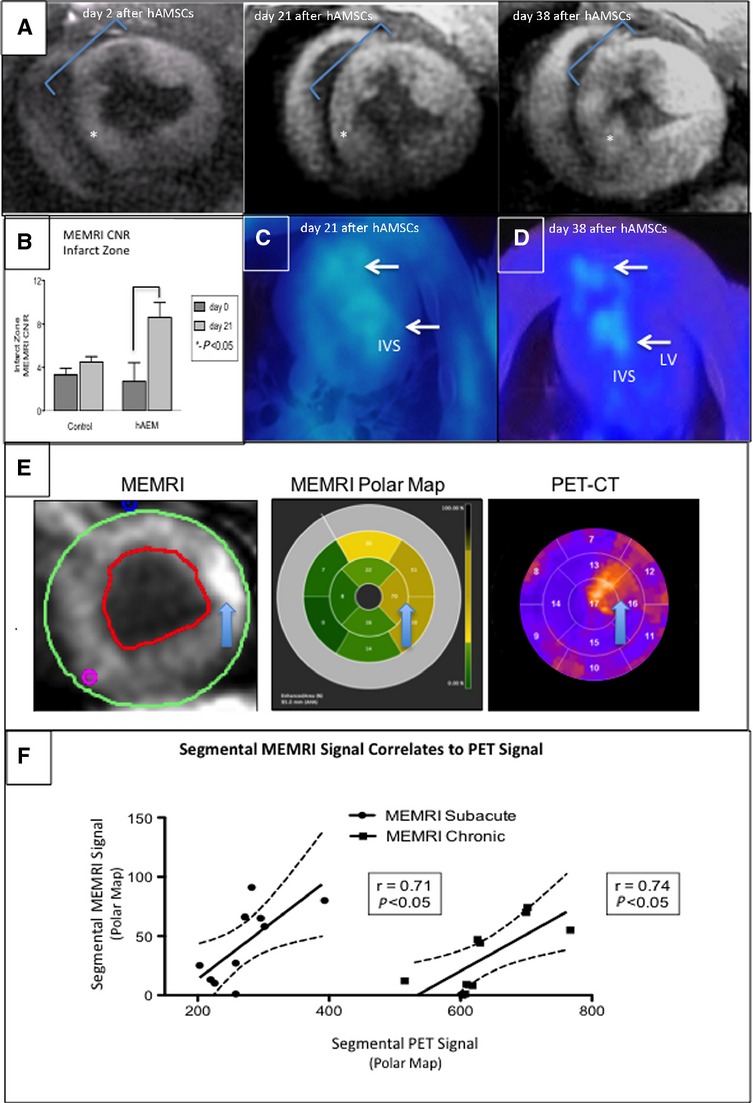
Figure 10.

MEMRI and PET colocalization of the hAMSC-injected hearts. In vivo confirmation of MEMRI signal for live hAMSCs within the myocardium of a hAMSC-injected swine is shown. hAMSCs were delivered 1 week after ischemia–reperfusion injury into the peri-infarct zones of the mid- to apical infero- and anteroseptum. A, SAX MEMRI image on day 2 with a characteristic MEMRI defect in the infarct zone (blue region of interest). Note area of inferoseptum with increased signal (star). Serial MEMRI shows increasing signal intensity in inferoseptum on days 21 and 38 after cell delivery, with overall increased signal within the infarct zone. B, Absolute increase in MEMRI CNR of the infarct zone in the hAMSC-treated vs control (normal saline–injected) hearts on day 21 after hAMSC delivery. C and D, Axial PET reporter gene signals from hAMSC populations in the apical septum, confirming specific live hAMSC activity on days 21 and 38 (IVS, LV). E, SAX MEMRI images (left) from an early hAMSC animal with traced endo- and epicardial contours; corresponding polar maps (middle) of MEMRI signal from the mid- to apical slices of the LV (the regions of hAMSC injection), with yellow and tan colors indicating increasing signal intensity; polar maps (right) from corresponding PET images. Note the similar patterns between MEMRI and PET polar maps. Arrows denote the focal signal in both MEMRI and PET images. F, Significant linear correlation of MEMRI signals (y-axis, signal intensity units) and PET signals (x-axis, signal intensity units) for both early hAMSC (left plot) and late-hAMSC (right plot) hearts. CNR indicates contrast-to-noise ratio; CT, computed tomography; hAMSCs, human amniotic mesenchymal stem cells; IVS, interventricular septum; LV, left ventricle; MEMRI, manganese-enhanced magnetic resonance imaging; PET, positron emission tomography; SAX, short-axis.
