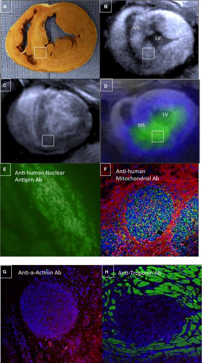
Figure 11.

Immunostaining confirmed intact hAMSC populations in vivo. A, Gross SAX section of a hAMSC-injected heart (6 weeks after cell delivery) demonstrated prominent anterior and inferoseptal peri-infarct segments, which were hAMSC injection sites (note: inset white box corresponds to MEMRI, DEMRI, and PET images and to the immunostaining tissue specimen). B, A bright focus (white box) of MEMRI signal is detected within the inferoseptal peri-infarct region corresponding to cell injection site. C, Matched DEMRI SAX image shows preserved myocardium (null signal) throughout most of the septum. D, 3-dimensional coregistration of PET-RG and MEMRI images acquired on the same day, showing a colocalized focus of intense inferoseptal signal from the PET-RG–transduced hAMSCs and increased MEMRI signal (white box). E and F, Myocardial sections stain positive for (E) anti-human nuclear antigen Ab and (F) anti-human mitochondrial antigen Ab, confirming the presence of live hAMSC populations up to 6 weeks after cell transplantation in these matched MEMRI and PET-positive regions. G and H, These same cell clusters are negative for both anti-α-actinin and anti-troponin Ab staining. Ab indicates antibody; DEMRI, delayed gadolinium enhancement magnetic resonance imaging; hAMSC, human amniotic mesenchymal stem cell; IVS, interventricular septum; LV, left ventricle; MEMRI, manganese-enhanced magnetic resonance imaging; PET, positron emission tomography; PET-RG, positron emission tomography reporter gene; SAX, short-axis.
