Figure 6.
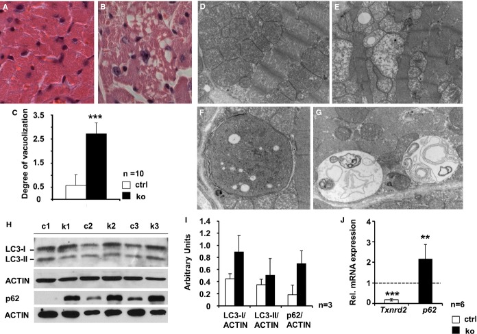
Autophagy in hearts of old Txnrd2−/− mice. Representative transverse sections obtained from old control (A and D), and knockout hearts (B, E, F and G). A and B, paraffin sections stained with hematoxylin and eosin. Note the markedly vacuolated cytoplasm in the knockout (B). C, quantification of vacuolization. D through G, Electron micrographs demonstrating severe mitochondrial degeneration in knockouts (E). Presence of double-membrane autophagosomes (F) and autolysosomes containing cellular material (G) in knockout cardiomyocytes (n=2). H and I, Western blots using protein homogenates obtained from 3 control (c1 to c3) and 3 knockout (k1 to k3) hearts of old mice probed with anti-LC3 and anti-p62 antibodies and corresponding quantification of protein expression. ACTIN was used as standard. J, Increased p62mRNA expression as analyzed by real-time RT-PCR, relative to control (set as 1). mRNA expression was normalized to Actin. Txnrd2-specific primers were used as internal control (P<0.01, n=6). Magnification: A and B, ×20; D and E, ×12 600; F and G, ×20 000. **P<0.01, ***P<0.001. LC3 indicates microtubule-associated protein 1A/1B-light chain 3; RT-PCR; real-time polymerase chain reaction; Txnrd2, thioredoxin reductase 2.

