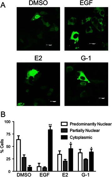
Fig. 2.

Estrogen activation leads to FOXO3a translocation. a Representative images of MCF7 cells transfected with FOXO3-GFP, starved of serum for 24 h and treated with 0.1 % DMSO, 50 ng/ml EGF, 50 nM estrogen, or 100 nM G-1 for 15 min. b Quantitation of the localization pattern of FOXO3-GFP-expressing cells from (a) as defined in Fig. 1. Results are reported as mean +/− s.e.m. from at least 3 experiments. *, p < 0.05; **, p < 0.01 vs. cytoplasmic localization of the DMSO control
