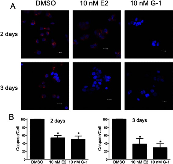
Fig. 7.

Estrogen and G-1 stimulation of MCF7 cells reduces caspase activation. MCF7 cells, under serum-free conditions, were treated with 10 nM estrogen or G-1 for either 2 or 3 days as indicated. Following treatment, cells were evaluated for caspase 7 activation employing a fluorogenic caspase substrate. a Representative images of each treatment at the indicated time point. Accumulation of the fluorescent product (red) is a result of caspase activation; blue, TO-PRO-3 staining of nuclei. b Images were analyzed for average fluorescence intensity on a per cell basis, and normalized to the intensity of the DMSO control. *, p < 0.05 relative to DMSO control
